
Description
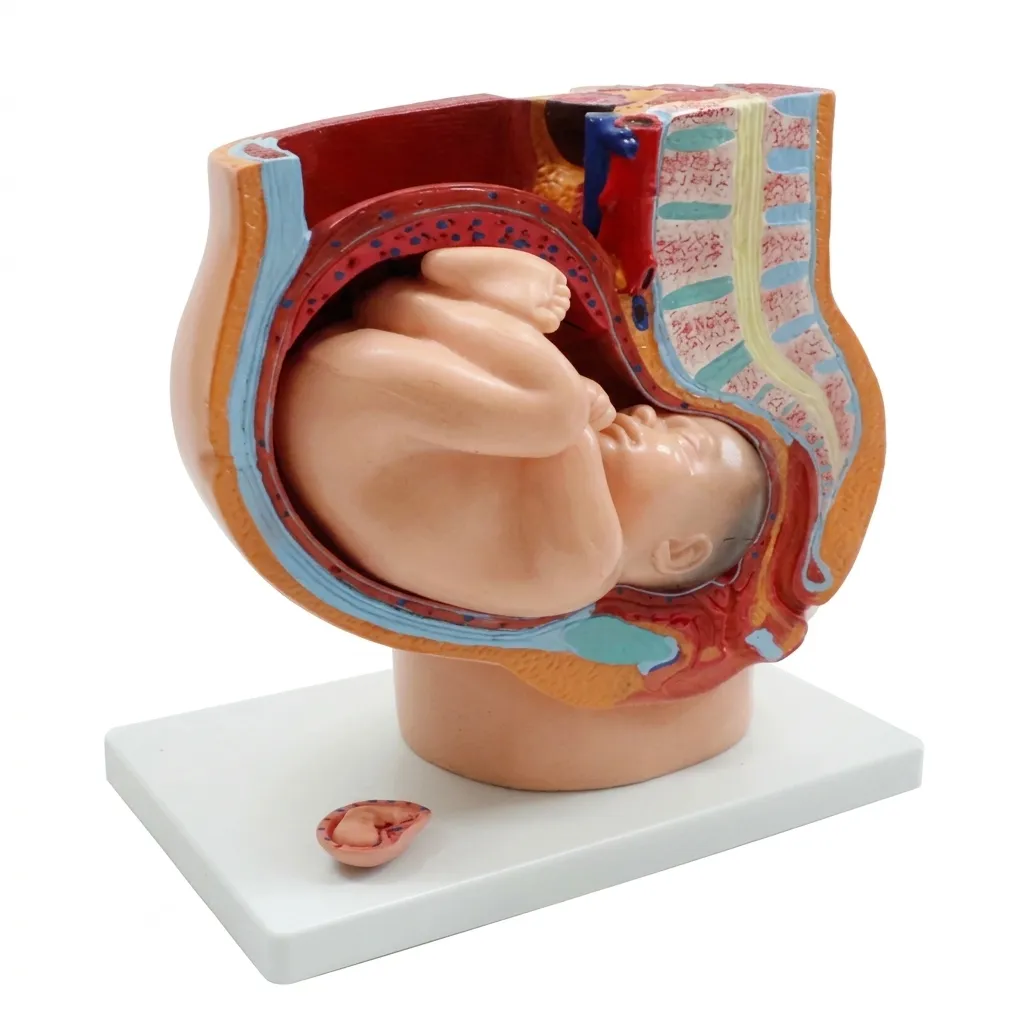
The XC-332B is a specialized, life-size anatomical model designed to illustrate the female reproductive system during the final stage of gestation. This median section model provides a comprehensive view of the anatomical changes and spatial relationships within the pelvic cavity during the 40th week of pregnancy.
Key Product Features
-
Full-Term Pregnancy View: Specifically depicts the female pelvis during the 9th month (40th week) of pregnancy, showing the normal position of the child before birth.
-
Removable Fetus: Includes a detachable full-term fetus, allowing for the study of fetal orientation, the umbilical cord, and the placenta.
-
Additional Embryo Stage: Uniquely features a smaller 3rd-month embryo mounted on the base for comparative study of early vs. late-stage development.
-
Detailed Pelvic Anatomy: The median section provides a clear 3D view of:
-
Reproductive Organs: The expanded uterus, cervix, and vaginal canal.
-
Internal Systems: The relationship between the compressed urinary bladder and the rectum.
-
Muscular Structure: Detailed representation of the abdominal and pelvic floor muscles.
-
-
High-Contrast Detailing: Hand-painted to differentiate between vascular structures, mucosal linings, and the muscular walls of the uterus.
Technical Specifications
| Feature | Details |
| Model Number | XC-332B |
| Scale | 1:1 Life-Size |
| Material | Advanced, non-toxic PVC |
| Parts | 4-part modular assembly |
| Dimensions | Approx. 38 x 25 x 40 cm |
| Mounting | Secured on a stable, professional display base |
Ideal For:
-
Obstetricians & Midwives: A perfect tool for explaining fetal presentation (vertex vs. breech) and the mechanics of the birth canal to expectant parents.
-
Medical & Nursing Students: Provides the spatial context needed to understand how a full-term pregnancy affects the surrounding internal organs.
-
Childbirth Educators & Doulas: An engaging visual aid for demonstrating the physical stages of labor and the descent of the fetus.
-
Biology & Anatomy Labs: Serves as a centerpiece for reproductive system studies and human development curriculum.
Why Choose the XC-332B?
The XC-332B stands out by offering both late-stage and early-stage context in one model. By including the 3rd-month embryo alongside the 40-week fetus, it allows for a powerful visual comparison of growth. Its 4-part disassembly ensures that educators can demonstrate the “internal” environment of the womb with the clarity and professional accuracy required for medical training.



Reviews
There are no reviews yet.